Search
Connect With us
 
Home Research Anti Microbial Activity
Anti Microbial Activity

        Antibacterial activity of Ficus racemosa Linn. Leaves on Actinomyces
               
- by Tanvir Shaikh*1, Ruksana Rub2, Kiran Bhise2, Pimprikar R.B.1, sufiyan A.


Abstract

The present study was undertaken with an objective to find out the antibacterial activity of Ficus Recemosa Linn. Leaves against Actinomycets viscosus .The hydro alcoholic extract of Ficus Recemosa Linn. Was found effective against Actinomyces vicosus (MTCC 7345). The minimum inhibitory concentration (MIC) was determined using Broth dilution technique and found to be 0.08mg/ml. The zone of inhibition was measured using Cup plate diffusion technique.

Key Words: Antibacterial activity, Ficus Recemosa, Actinomyces viscosus, leaves.

Back to Top
Introduction
Ficus Recemosa Linn is a large dedicious tree distributed throughout India particularly in evergreen forests and moist localities11.Root bark, leaves fruit and galls are part of tree used for therapeutic activity. Bark, leaves and unripe fruit are carminative, astringent, stomachic and vermicide. As mentioned in the Ayurvedic Nighanthus that the infusion of the bark, fruit and leaves is cooling, sweet and astringent2. The bark chemically constitutes of gluanol acetate, beta-sitosterol, leucocynedin and leaf chemically contain beta-amyrin, beta-sitosterol and tannin. Fruit chemically contain lupeol-OAc, glucose, sterol, and gluanol-OAc3-5. Actinomyces viscosus belongs to group of actinomycets. It is gram positive, aerobic, non sporing, rod shaped bacteria. The mouths contain a wide variety of bacteria, but only a few specific species of bacteria are believed to dental cares. For root caries the most closely associated bacteria frequently identified are Actinomyces viscosus, Nocardia spp and streptococcus mutans. Actinomyces viscosus is frequently encountered in high proportion of smooth tooth surface and gingiva6-9. Antibacterial Tests are indicated for those organisms contributing to the infectious process whose susceptibility can not be predicted from knowledge of their identity. The Antibacterial activity has been done by two methods. Dilution method is used to determine the minimal concentration usually expressed
in units or microorganism per ml of an antimicrobial agent required to inhibit or kill a microorganism. Diffusion method is used to test the bacterial susceptibility to antimicrobial agent and may be measured in-vitro by using the principles of agar diffusion10-11.
Back to Top
Materials and Methods
Plant material:
The fresh leaves of the Ficus Recemosa Linn belong to family Moraceae were collected from different places of Bhavani peth, pune. The leaves of Ficus Recemosa Linn were identified and authenticated from ‘Agarkar Institute’ pune.

Preparation of Extract:
A coarse powder of the leaves of Ficus Recemosa Linn was prepared and dried at 500c. The coarse powder extracted using hydroalcohoic (methanol:water) in soxhlet apparatus. The extracts were then subjected to photochemical screening using standard procedure.

Test Organism:
The extract were screened against bacteria i.e. Actinomyces Viscosus (MTCC 7345).

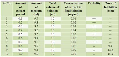
Table 1:
Showing protocol for evaluations of MIC, results of Broth Dilution technique and Cup Plate Diffusion.

++ Turbidity present, -- Turbidity absent.

Antibacterial Activity:

A) Broth Dilution technique:
Preparation of extracts of Ficus racemosa leaves: 0.1 gram (100mg) of dried evaporated extract was dissolved in 100ml of 70% methanol giving final concentration of 1 mg/ml.

Preparation of sterilizing media :
The microbial work was carried out in aseptic area. Brain Heart Infusion broth was prepared. The medium was poured in the tubes which were then sterilized by autoclave using 15 lb pressure at 1210C for 15 minutes. Using sterile pipettes exact amount of extract was added as indicated in the Table 1 to obtain a final volume of 10ml. The tubes were then inoculated with 0.05ml of the standardized culture. The tubes were incubated at temperature 300C for 48 hours. The tubes were observed for growth of microorganism by observing the turbidity produced. The test procedure was repeated three times to check the reproducibility of the results. The lowest concentration that inhibits the growth is the Minimum Inhibitory Concentration (MIC). Penicillin was used as reference standard.

B) Cup Plate Diffusion Method:
All the glassware and the Petri plates were sterilized by dry heat in an oven at 1600C for one hour. Brain Heart Infusion agar was prepared in distilled water. Molten agar was poured in each test tube and plugged with non absorbent cotton. All the test tubes were sterilized by autoclaving using 15 lbs pressure at 1210C for 15 minutes. The molten agar
was poured in sterile Petri plates aseptically and allowed to solidify at room temperature. All the Petri plates were aseptically flooded with 0.1ml of the standardized culture. The holes of 7mm were bored aseptically using sterile cork borer. The agar plugs were taken out carefully so as not to disturb the surrounding medium. The holes were filled completely with desired extract and kept in incubator at 300C for 48 hours. After this the Petri plates were
observed for the antibacterial activity and zone of inhibition was measured. The solvent effect was neutralized.

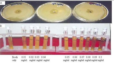
Figure 1: Showing results of Cup Plate Diffusion method and Broth Dilution method.
A: Showing results of Cup Plate Diffusion Method
B: Showing results of Broth Dilution Method


Back to Top
Results & Discussion

Determination of Minimum Inhibitory Concentration for antibacterial activity

A. Broth Dilution technique :
The tubes were incubated at temperature 300C for 48 hours. The tubes were observed for growth of microorganism by observing the turbidity produced. The tubes having concentration of extract in final solution from 0.01 mg/ml to 0.07 mg/ml shown the presence of turbidity when incubated at temperature 300C for 48 hours. The tubes having concentration of extract in final solution from 0.08 mg/ml to 0.1 mg/ml produced clear solutions when incubated at temperature 300C for 48 hours. The test procedure was repeated three times to check the reproducibility of the results. The lowest concentration that inhibited the growth of microorganism Actinomyces viscosus was 0.08 mg/ml. From this data Minimum Inhibitory Concentration of the extracts of Ficus racemosa leaves for microorganism Actinomyces viscosus was found to be 0.08 mg/ml.

B. Cup Plate Diffusion Method :
The Petri plates were incubated at temperature 300C for 48 hours. When the Petri plates were observed for the antibacterial activity and zone of inhibition was measured The Petri plates having concentration of extract 0.07 mg/ml incubated at temperature 300C for 48 hours did not show any zone of inhibition. The Petri plates having concentration of extract 0.08 mg/ml to 0.1mg/ml incubated at temperature 300C for 48 hours produced zone of inhibition. Metronidazole gel was used as positive control produced zone of inhibition. The test procedure was repeated three times to check the reproducibility of the results. The concentration of extract of Ficus racemosa leaves 0.08 mg/ml, 0.09 mg/ml; 0.1 mg/ml shown the zone of inhibition.The extract of Ficus racemosa leaves of 0.08mg/ml to 0.1 mg/ml have better antibacterial acticity.

Back to Top
References
[1] The Ayurvedic Pharmacopoeia of India, Government of India, Ministry of Health and Family Welfare, Department of Indian System of Medicine and Homeopathy, New Delhi.Ist Edition, Part I, Vol. I, 117-118, (1990)

[2] Dr.K.M.Nadkarni’s,revised by Dr.A.K.Nadkarni, Indian Materia Medica,Bombay Popular Prakashan, Mumbai, IInd reprint IIIrd revised edition, 548-550,(2000)

[3] C.P.Khare, Indian Medicinal Plants An Illustrated Dictionary, Springer (India) Private Limited, New Delhi, Ist Indian reprint,266-267,(2007)

[4] The Wealth Of India, A Dictionary Of Indian Raw Materials and Industrial Products (Raw Materials) National Institute Of Science Communication,Council of Scientific and Industrial Reseach, New Delhi, Ist Supp.Set.Vol III, 130, (2002)

[5] The Wealth Of India, A Dictionary Of Indian Raw Materials and Industrial Products (Raw Materials) National Institute Of Science Communication,Council of Scientific and Industrial Reseach, New Delhi, Ist Reprint.Set.Vol IV, 35, (2002)

[6] William J,Hausler Jr MS,Topley & Wilson’s Microbiology µbial infectionsActinomyses, Actinobacillosis and releated diseases( Great Britan – Arnold) Vol.3 9th ed. 777-787,(1998).

[7] R.Ananthanarayan,C K J Panikar,Textbook of Microbiology, Orient Longman Limited,Madras,6th ed.370-373,(1992)

[8] J.P. Duguid, B.P. Marmion, R. H. A. Swain, MACKIE & McCARTNEY Medical Microbiology, Vol 1,microbial infections, 13 th ed. Churchill Livingstone,304,(1980)

[9] Michael J. Pelczar,JR., E.S.C. Chan, Noel. R. Krieg, Microbiology,TataMcGraw-Hill publishing, 5th ed. 274-275,(1997)

[10] B.J Wadher, G.L Bhoosreddy, ‘Manuals of Diagnostic Microbiology’, Himalaya Publishing House,IST edition, 62- 67,(1995).

[11] Barry A.L.,The antimicrobial susceptibility test principles and practices, Lea and Febiger, Philadelphia,163-164,(1976)

Back to Top
Research
 
 
Photo Gallary
Contact us
Address :Rahul Apartment, Satbhai Lane, Dilhi gate, Ahmednagar - 414001
Ph.no.: (0241)2345894
Email: nkortikar@gmail.com